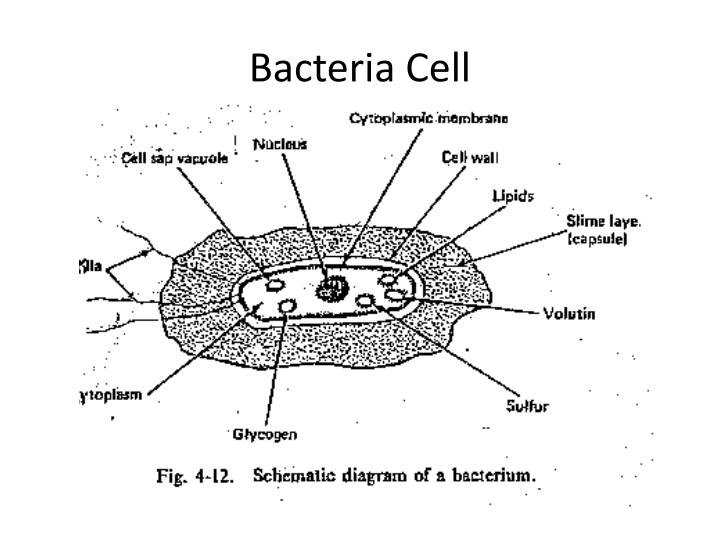
39 bacterial cell picture with labels
3 Common Bacteria Shapes - ThoughtCo The three basic shapes of bacteria include cocci (blue), bacilli (green), and spirochetes (red). Bacteria are single-celled, prokaryotic organisms that come in different shapes. They are microscopic in size and lack membrane-bound organelles as do eukaryotic cells, such as animal cells and plant cells. Bacteria are able to live and thrive in ... Molecular Expressions Cell Biology: Bacteria Cell Structure In gram-negative bacteria, the cell wall is thin and releases the dye readily when washed with an alcohol or acetone solution. Cytoplasm - The cytoplasm, or protoplasm, of bacterial cells is where the functions for cell growth, metabolism, and replication are carried out. It is a gel-like matrix composed of water, enzymes, nutrients, wastes ...
Bacteria Diagram Labeled | Cell diagram, Prokaryotic cell, Medical ... 445 views. Find this Pin and more on Biology by Fernanda Newman. Cell Structure. Cell Wall. Korean Words. Science Facts. Microbiology. Study Notes. Medical School.

Bacterial cell picture with labels
Bacteria cell diagram Images, Stock Photos & Vectors - Shutterstock 2,581 bacteria cell diagram stock photos, vectors, and illustrations are available royalty-free. ... Find Bacteria cell diagram stock images in HD and millions of other royalty-free stock photos, illustrations and vectors in the Shutterstock collection. Thousands of new, high-quality pictures added every day. ... Image Library | CDC Online Newsroom | CDC Under a high magnification of 21674X, this digitally-colorized, scanning electron microscopic (SEM) image depicts a view of a dividing, Escherichia coli bacterium, clearly displaying the point at which the bacteria's cell wall was splitting into two separate organisms. See PHIL 7137 for a black and white version of this image. Bacteria in Photos - photo gallery of bacteria facultatively anaerobic bacteria: Motility: nonmotile: Catalase test: catalase-positive: Oxidase test: negative* Spores: non-spore forming * Some species (non-human isolates) are positive: Streptococcus: ... (the cells stain a weak Gram-negative) Microscopic appearance: Spirochetes: Oxygen relationship: microaerobic: Motility: motile: Catalase ...
Bacterial cell picture with labels. Bacteria Labeled Stock Illustrations - Dreamstime New users enjoy 60% OFF. 184,305,164 stock photos online. Download 220 Bacteria Labeled Stock Illustrations, Vectors & Clipart for FREE or amazingly low rates! New users enjoy 60% OFF. 184,305,164 stock photos online. ... Coronavirus bacteria cell labeled , 2019-nKoV New coronavirus bacteria. No infections and stop coronavirus concepts ... Bacteria Under Microscope Stock Photos and Images - Alamy Find the perfect bacteria under microscope stock photo. Huge collection, amazing choice, 100+ million high quality, affordable RF and RM images. ... Microbes and viruses of different shapes and colors isolate. Bacterial cells, m Editable vector illustration of bacteria under a ... Search Results for Bacteria Under Microscope Stock Photos and ... Animal Cell Labeled Pictures, Images and Stock Photos Browse 116 animal cell labeled stock photos and images available, or start a new search to explore more stock photos and images. Newest results. Diagrams of animal and plant cells. Labelled diagrams of typical animal and plant cells with editable layers. Components of Eukaryotic cell, nucleus and organelles and plasma... BYJUS The structure of bacteria is known for its simple body design. Bacteria are single-celled microorganisms with the absence of the nucleus and other c ell organelles; hence, they are classified as prokaryotic organisms. They are also very versatile organisms, surviving in extremely inhospitable conditions. Such organisms are called extremophiles.
Draw a labelled diagram of a bacterial cell. - Careers360 Buy Now NEET Foundation + Knockout NEET 2024 (Easy Installment) Personalized AI Tutor and Adaptive Time Table, Self Study Material, Unlimited Mock Tests and Personalized Analysis Reports, 24x7 Doubt Chat Support,. Animal Cell Labeled Diagram Pictures, Images and Stock Photos Browse 19 animal cell labeled diagram stock photos and images available, or start a new search to explore more stock photos and images. Newest results. ... Labeled educational bacteria internal structure scheme. Biological blue green algae diagram with carboxysome, thylakoid and phycobilisome parts location inside cell. animal cell labeled ... Label the Bacterium Cell - EnchantedLearning.com The cell is the basic unit of life. The following is a glossary of Bacterium cell terms. basal body - A structure that anchors the base of the flagellum and allows it to rotate. capsule - A layer on the outside of the cell wall. Most but not all bacteria have a capsule. cell wall - A thin membrane located outside the plasma membrane and within ... A Labeled Diagram of the Animal Cell and its Organelles A Labeled Diagram of the Animal Cell and its Organelles. There are two types of cells - Prokaryotic and Eucaryotic. Eukaryotic cells are larger, more complex, and have evolved more recently than prokaryotes. Where, prokaryotes are just bacteria and archaea, eukaryotes are literally everything else. From amoebae to earthworms to mushrooms, grass ...
Bacteria Labeled Diagram Stock Vector Image & Art - Alamy Download this stock vector: Bacteria Labeled Diagram - EG0XT7 from Alamy's library of millions of high resolution stock photos, illustrations and vectors. Animal Cell - Free printable to label + Color -kidCourses.com GOLGI BODIES modifies, stores, sorts & secretes the cells chemical products. LYSOSOMES responsible for intracellular digestion. CYTOPLASM the semi-fluid interior part of the cell. VACUOLE "bubble" for storage. CENTRIOLES help with cell division. Animal Cell for Kids - Label the Parts and Color! Below is the answer key: Structure of Bacteria (With Diagram) | Microbiology 8. According to Peberdy (1980) the only compound present in the cell walls of both Gram-negative and Gram-positive bacteria is 'peptidoglycan'. The cell walls of Gram-positive bacteria contain up to 95% peptidoglycan and up to 10% teichoic acids. 9. Cytoplasmic membrane is a thin (5-10 nm) layer lining the inner surface of the cell wall. Structure of Bacterial Cell (With Diagram) - Biology Discussion Cell wall: It is a tough and rigid structure of peptidoglycan with accessory specific materials (e.g. LPS, teichoic acid etc.) surrounding the bacterium like a shell and lies external to the cytoplasmic membrane. It is 10-25 nm in thickness. It gives shape to the cell. Nucleus: The single circular double-stranded chromosome is the bacterial genome.
cell_poster_bacteria_types.JPG 1,182×1,555 pixels | LAB | Pinterest | Poster
Bacteria in Microbiology - shapes, structure and diagram Bacteria shapes, structure and diagram. by Razi Brown. Bacteria cells are the smallest living cells that are known; even though viruses are smaller than bacteria, viruses are not living cells. There are different types of bacteria with various sizes, shapes, and structures. The bacteria shapes, structure, and labeled diagrams are discussed below.

Fun With Microbiology (What's Buggin' You?): Bacterial Vaginosis
97,783 Bacteria Cell Stock Photos and Images - 123RF Bacteria Cell Stock Photos And Images. 97,783 matches. Page of 978. Structure of a bacterial cell. Anatomy of the prokaryote. unicellular organism. Vector diagram for your design, educational, medical, biological and science use. Bacteria vector icon isolated on transparent background, Bacteria logo concept.
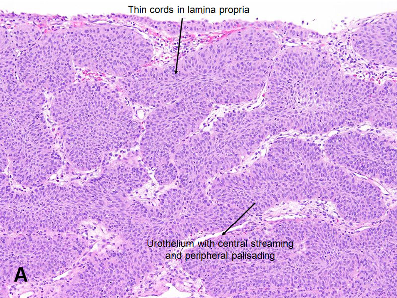
American Urological Association - Inverted Papilloma
Bacterial Cell Structure Labeling Diagram - Quizlet Bacterial Microcompartment. Protein coated packets used to localize enzymes and other proteins into the cytoplasm. Plasmid. Double-stranded DNA circle containing extra genes. Flagella. specialized appendage attached to the cell by a basal body that holds a long rotating filament. Pushes cell forward. Endospore.
Review
Bacteria Cell Structures with Labels Stock Vector - Dreamstime Bacteria Cell Structures with labels. Illustration about infection, bacilli, include, bacteriology, labeled, antibacterial, morphology, capsule, diplococci, anatomy - 186260912 ... Affiliate / Reseller Upload & sell photos. Dreamstime Facebook Dreamstime Twitter Dreamstime Pinterest Dreamstime Instagram Dreamstime Linkedin Dreamstime YouTube.
PPT - Wastewater Microbiology PowerPoint Presentation - ID:1635113
Plant and Animal Cells - Labeled Graphics A compilation of plant and animal cell images with organelles and major structures labeled. Students can print images to help them learn the cell. ... if students missed the lab that day they can view a site with pictures to complete lab handout Plant Cell ... looks at cheek and onion cells. Prokaryote Coloring - color a typical bacteria cell ...
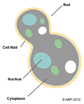
Fungi - ABPI - Resources for Schools
Interactive Bacteria Cell Model - CELLS alive Pili, Fimbriae: These hollow, hairlike structures made of protein allow bacteria to attach to other cells. A specialized pilus, the sex pilus, allows the transfer of plasmid DNA from one bacterial cell to another. Pili (sing., pilus) are also called fimbriae (sing., fimbria). Flagella: The purpose of flagella (sing., flagellum) is motility.

PHARMA WISDOM: Mechanism of Action of Chemotherapeutic drugs
Structure of a bacterial cell, labeled. Stock Illustration Download Structure of a bacterial cell, labeled. Stock Illustration and explore similar illustrations at Adobe Stock. Adobe Stock. Photos Illustrations Vectors Videos Audio Templates Free Premium Editorial Fonts. Plugins. 3D. Photos Illustrations Vectors Videos Audio Templates Free Premium Editorial Fonts.

Pathology Outlines - Plasma cells
Bacterial cells - Cell structure - Edexcel - GCSE Combined Science ... Feature Eukaryotic cell (plant and animal cell) Prokaryotic cell (bacterial cell) Size: Most are 5 μm - 100 μm: Most are 0.2 μm - 2.0 μm: Outer layers of cell

Katy J Negus. BA Hons. CG Arts & Animation: Making an Antibody Research
50 Striking Microscopic Images of Viruses and Bacteria Click through the slideshow above to see 50 striking electron micrographs of some of the world's most dangerous and deadly disease-causing viruses and bacteria. Know your flu risk. Check out the ...
Post a Comment for "39 bacterial cell picture with labels"